Heart dissection cont.
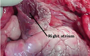
Why
are the walls of the atrium so thin when compared to those of the ventricle?
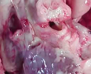
The
picture on the left shows the vena cava (major vein) on the left and
the aorta on the right. Note the relative thickness of each vessel.
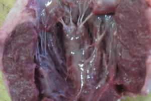
- interventricular septum,
- papillary muscles,
- chordae tendinae,
- right ventricular wall,
- left ventricualr wall,
- tricuspid valve,
- mitral valve (bicuspid),
- trabeculae carneae.
- moderator band
Compare the difference in thickness between the left and right ventricles. Explain why there is such a difference.