
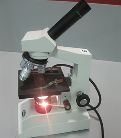
A most familiar piece of equipment in a biology laboratory is the compound microscope. This instrument uses lenses and light to enlarge images. Each compound microscope has two systems of lenses for greater magnification, the ocular, or eyepiece lens and the objective lens, or the lens closest to the object. Click to see the parts of the compound microscope.
One detail that is provided by the microscope and is important when describing the specimen is the magnification. Magnification of an object is obtained by multiplying the magnification of the eye piece with the magnification of the objective lens. For example, if the eye piece is 10X and the objective lens is 20X then the specimen is magnified 200 times.
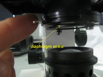
By manipulating this lever and controlling the amount of light coming through one can achieve better resolution.
The video on the right shows how the image changes as we change the amount of light coming through by adjusting the iris.
Notice how detail is lost when too much or not enough light is allowed through.
After preparing the slide and placing it on the stage it is time to focus the specimen. The proper way to focus a microscope is to start with the lowest power objective lens first and while looking from the side, turn the coarse focus knob so the objective lens is as close to the specimen as possible without touching it. Now, looking through the eyepiece lens, turn the coarse knob so that the objective lens move up until the image is in focus.
When viewing a specimen under high magnification you can observe greater detail if you manipulate the fine focus to observe detail of organelles that may be deeper inside the organism. This is known as the depth of field and the greater the magnification the shallower is the depth of field.
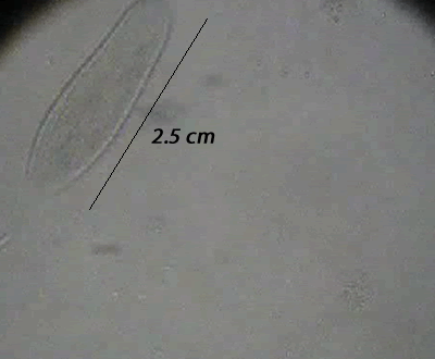
Take the image of the spirogyra, pictured on the right. By manipulating the fine focus we can see detail of the chloroplasts and cell wall deeper inside the cell.
Another term that you will need to know when working with microscopes is the "Field of view". This is the amount of the specimen that can be seen through the eye piece. The field of view decreases as the magnification increases.
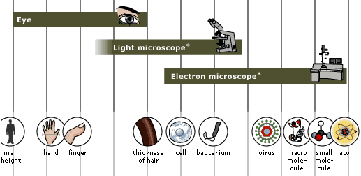
The image on the left shows the different objects that can be resolved with a light microscope and electron microscope.
a) A molecule of the protein known as collagen can be seen using an
b) A virus particle can be seen using an
c) A Human red blood cell can be seen using an
d) A protein molecule that acts as a hormone receptor attached to the cell membrane can be seen using an