The kidneys are the filtering
units of the body. They purify the blood by first filtering it and then
reabsorbing those molecules that are important for a healthy body. Dissecting
a kidney reveals the many subtle features of the body's filtering system.
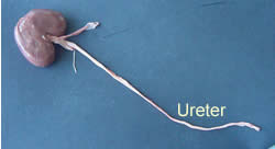
Looking at a fresh sheep's kidney we can identify three features. The ureter, renal vein and renal artery.
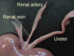
The renal artery can be identified as the thicker of the other two tubes coming from the kidney.
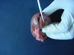
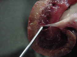
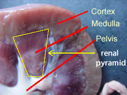
Once the kidney is cut lengthwise, three regions can be identified.
The cortex is the outer dark, red-brown layer, while the medulla is the lighter layer just beneath the cortex. The pelvis is the end of the ureter that flares out in all directions. The pelvis is really a funnel like, muscular, cavity that collects urine. Smooth muscle fibers line the wall of the pelvis and allow for a movement of urine into the ureter.
Structures such as the minor calyx and the major calyx can also be seen and provide channels through which urine flows from the collecting tubules to the ureter.
The image on the right shows how the different coloured areas accommodate the nephrons. Notice how the nephrons are arranged amongst the different regions of the kidney. Bowman's capsules are found in the cortex and it is there that filtration of the blood takes place. Twenty percent of the fluid in the blood passes through the porous holes of the glomerulus and moves into Bowman's capsule.
Two types of nephrons exist. One has a short loop of Henle and is known as a cortical nephron the other has a longer loop of Henle and is known as a juxtamedullary nephron.
Looking at the image on the right answer the following questions.
Both type of nephrons have their Bowman's capsules in which region?
In which region is the loop of Henle?
What type of filtrate is produced by the juxtamedullary nephrons?