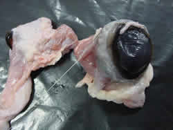
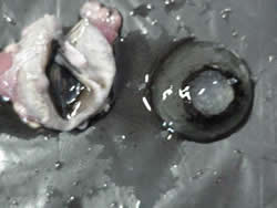
Squeeze the contents out.
View the video on the right. You will see black particles floating in a thick jelly-like medium (vitreous humor). These black fragments are parts of the ciliary muscles that have dislodged in the process of dissecting the eye.
What is the function of the vitreous humor? (research task)
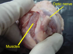
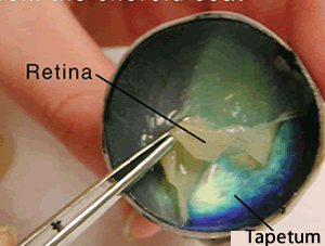
Most of the light sensitive tissue, retina, is not attached to the eye. Instead, it is held in place by the dense fluid known as the vitreous humor. You will most probably not see the retina as it will peel away from the back of the eye easily. If you do manage to find the retina you will see that it gathers at the back of the eye at a point known as the blind spot or optic disc. It is there that nerve fibers from each nerve cell forming the retina move out or the eye via the optic nerve.
If you do see the retina use a pair of tweezers to gently lift it from the inside wall of the eye. It will most likely tear as it is a very delicate structure. Beneath the retina you will see a shiny and colorful tissue called the tapetum lucidum. This layer is important in night vision and helps animals see greater detail in low light. Humans do not have such a structure in their eye but animals such as cats and crocodiles do and is the reason high their eyes reflect in the dark when a torch is directed onto their eyes. By reflecting light back through the retina the retina can process images in low light conditions.
Sourced from https://www.youtube.com/watch?v=5nGZvonGrYw at 11.39am 10/10/20
The lens along with the vitreous humor are expelled as shown on the left.
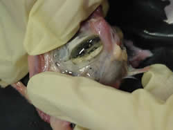
When removing the lens take care not to damage it. Hold it up at eye level and view a distant object through the lens.
Describe the image you see.
You will notice it is like a magnifying glass.
Pass the lens over a page of writing.
Why do you think the vitreous humor is so thick?
Take the lens and look through it at a distant object. Describe the image you see.
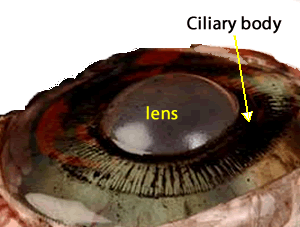
Have a look at the inside surface of the eye. You will notice the black surface inside the eye. Apart from the tapetum lucidum why do you think the internal surface of the eye is black?
Why is it important, if the eye is punctured by an object, such as a stick or nail,
that we do not pull it out but keep it in place and go to a hospital immediately?
A dark coloured pigment, called melanin, surrounds the tapetum lucidum. Its job is to absorb any light that falls on it. This prevents uncontrolled reflection of light inside the eye that may cause confusing images to form.
Albinism is a condition where the body is unable to produce melanin. Albino humans have poor eyesight. Explain why.

You can simulate how the lens of the eye functions with a magnifying glass and piece of paper. The paper represents the retina while the magnifying glass represents the lens.
a) What can you say about the image formed by the lens on the retina?
b) What happens to the image as the distance from the paper to the magnifying glass is varied?
c) How does the lens manage to focus an image on the retina if it can not move?