Heart dissection
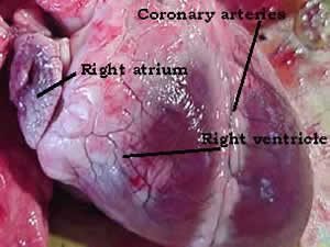
On the right is a picture of a sheep's heart before dissection.
Note the relative size of the atrium as compared to the ventricle.
Find out the role of the coronary blood vessels.
The image on the left shows a cross section through the left ventricle. Notice the thickness of the wall of the ventricle. The left ventricle pumps blood a long way round the body. It must generate high pressure in order to push the blood and this requires a strong muscular contraction.
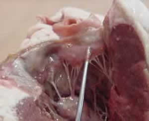
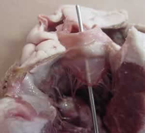
Notice the type of valves that exist in the heart. Look at the valves separating the atrium and ventricle. Describe their appearance and give an explanation of how they work. Click to see a 300 kb video of the working heart.
Between the right atrium and the right ventricle you will notice three clear membranes connected by tendons called chordae tendinae. The three membranes form the tricuspid valve and are connected via the chordae tendinae to a muscular structure bulging from wall of the ventricle known as papillary muscle. Papillary muscles exist in both the right and left ventricles, three in the right ventricle and two in the left ventricle. Just before ventricular contraction takes place the papillary muscles start to contract in both ventricles pulling on the chordae tendinae. This prevents blood flowing back into the atria. Valves serve to keep blood flowing in one direction as it passes through the heart.
Between the left atrium and left ventricle is a valve made of two membranous flaps known as the bicuspid valve or mitral valve. Much like the tricuspid valve the flaps will be connected to papillary muscles by chordae tendinae.
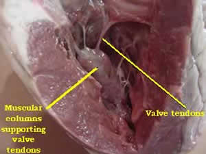
Using a probe, lift the flaps of the heart valve. Notice how the flaps are attached to the muscle by tendons. Why are the tendons necessary.
Valves become
diseased and the person's mobility is severely reduced.
How does a dysfunctional valve reduce the persons ability to exercise?
Artificial valves replace many diseased and damaged human valves. Pig
heart valves are commonly used to replace human valves. Knowing how real heart valves work, describe how an artificial valve works..
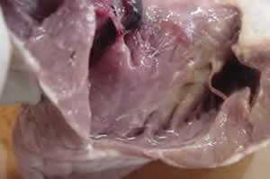
Using a probe investigate how the blood flows through the heart. The blood vessel that the probe is pushed through is an artery. Notice its thick elastic walls.
Why are the walls of arteries so thick and elastic?
Identify the veins and arteries on you heart specimen.
The right ventricle has walls that are thinner in muscle tissue than the walls of the left ventricle.
Why do you think this is so? Think of how far the blood has to travel when it leaves the right ventricle.
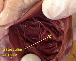
The interiror of the atrial and ventricular walls are lined with woven ridges of cardiac muscle. These ridges found in the ventricle are known as trabeculae carneae and those found int he atrium are known as pectinate muscle. These woven arrangement allows for a greater strength of contraction with a relatively smaller mass of muscle tissue.