Sports Science
The contraction
When a signal arrives at the neuromuscular junction a chemical called acetylcholine is ejected from the motor nerve ending and into the gap between the nerve and the muscle cell. Here it sits on special receptors and causes chemical changes to take place that initiate the contraction mechanism.
Inside
every muscle cell there are hundreds of long protein filaments called
myofibrils. When the contraction mechanism is
initiated the myofibrils become shorter hence contracting the cell. The
mechanism by which the myofibrils become shorter is called the "sliding
filament" model of muscle contraction.
Each
myofibril is a linear arrangement of sarcomeres.
The sarcomere is the basic contractile unit of muscle. A sarcomere is
pictured on the right.
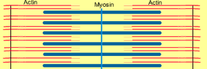
Within the sarcomere two proteins, myosin and actin, are precisely arranged resulting in the striated appearance of cardiac and skeletal muscle when viewed under a microscope.
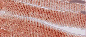
A picture of the striated appearance of skeletal muscle is shown on the right.
Energy,
in the form of adenosine triphosphate (ATP),
is required for the protein filaments to slide over one another. ATP is
required for both contraction and relaxation. This is why after death,
when the ATP is exhausted, the muscles go into a state of rigor mortis.
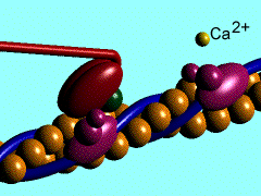
The exact mechanism is beyond the scope of this unit of study but the
animation below may prove helpful.
Myosin
grabs onto the actin filament and pulls it inwards. We will note a few
things from the animation.
- Ca2+ ions (calcium ions) are involved
- Mg2+ ions (magnesium ions) are also involved
- ATP attaches itself to the myosin head as it links onto the actin filament.
- ATP is converted into ADP with the release of a phosphate group (P). This conversion provides the energy for the contraction process.