The brain is composed of billions of nerve cells, all interconnected . Signals pass from one neuron to the other not by contact but over tiny gaps called synapses.
The brain is divided into many regions, all with highly specific functions and connected together for greater communication.
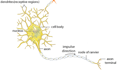
A basic type of nerve cell is called the Neuron. Neurons have excitable membranes that can depolarize to produce an action potential. They also have specialised extensions called dendrites, cell body, axon and terminal axons, as shown on the right
Different types of neurons exist.
-
sensory neurons - these are a diverse group of nerve cells that are specialised for detection of stimuli and the the transfer of the signal to the brain - examples include:
a) photoreceptors in retina of eye
b) olfactory neurons in nose
c) pressure sensitive neurons in skin
d) temperature sensitive neurons in skin
e) pain sensitive neurons everywhere
-
motor neurons - transmit signals from the brain to muscles or glands
- interneurons - neurons found in the brain that transfer signals from one nerve cell to another
Refer to the rubric before starting.
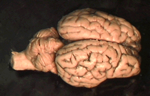
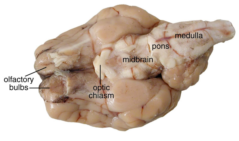
Draw a diagram of the exterior of the sheep's brain, as shown on the left. You will notice that surface of the cerebrum is organised into folds called gyri, separated by grooves called sulci.
On your diagram indicate the following:
- two cerebral hemispheres
-
frontal lobe
-
temporal lobe
-
occipital lobe
- parietal lobe
- corpus callosum (This is identified if you use your fingers to separate the cerebral hemispheres)
- cerebellum.
- pineal gland
- superior collicullus
- thalamus
Click to see a diagram that might help.
The cerebrum is deeply wrinkled and folded? Explain why.
What could be some of the effects of the folding? Think of communication between different areas of the brain.
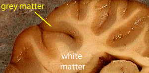
Cut a small cross-section of one of the cerebral hemispheres. You will notice a band of darker coloured tissue called grey matter. In this area exists neurons that do not have long axons. The nerve cells of the grey matter form connections with each other creating networks through which nerve signals travel. This is where cognitive processing occurs.
The dark coloured top layer appears darker because of the cells' dark nuclei. This is called the cortex and contains roughly 70% of the one hundred billion or so nerve cells in the brain.
The inner, lighter layer is called white matter. This tissue is composed of the long extensions that transmit a signal called the axons.
The white matter, is composed of neurons that have long myelinated axons, as shown above. It forms the structures at the center of the brain, like the thalamus and the hypothalamus. It is found between the brainstem and the cerebellum. It is this white matter that allows communication to and from grey matter areas, and between grey matter and the other parts of the body, such as muscles and major organs.
Axons are protected by the myelin sheath, which provides insulation from the electrical processes, allowing them to transmit nerve signals faster than unmeylinated axons. It is also the myelin that is responsible for the white appearance of the white matter. 60 percent of the brain is comprised of white matter. However 94% of the oxygen consumbed by the brain is used by the grey matter.
Why is the spinal cord composed of white matter?If myelin allows for faster conduction of signals why do the neurons of grey matter not have myelinated axons?
Describe the distribution of white and grey matter. Give a reason for this.
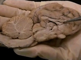
Using a scalpel, cut the brain in half as shown on the left. Label the following
- cerebellum
- corpus callosum
- ponds
- midbrain.
Indicate the location of the hypothalamus on your diagram. What is the purpose of the hypothalamus?
What is the function of the pituitary gland? (Research may be required to answer this question)
What is the function of such fluid filled chambers in the brain?
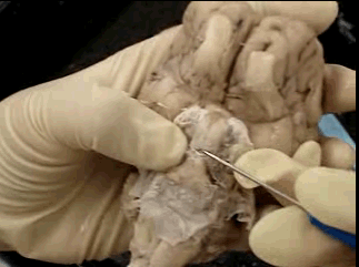
What is the name of the structure pointed to by the teacher in the image on the left?
What is its function
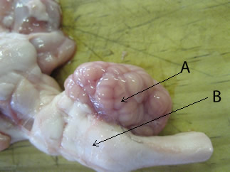
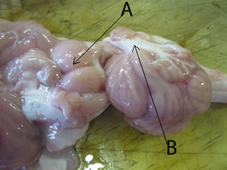
4) Identify the structures A and B
These slides may help.
View of the brain from beneath.
Cross section of the brain
The brain stem is made up of the pons, medulla and cerebellum. Identify the parts of the brainstem in your drawing or digital image.
5) i) Identify structure A
. What is its function?
ii) The white matter (B) is most likely
iii) When dissected, the white matter feels greasy and fatty. What possible explanation can you provide?